

What Is A Question That Could Be Answered By Observing Chromosomes Of Different Species Of Animals
7.8: Errors in Chromosome Number
- Page ID
- 35704
What you'll larn to practise: Examine karyotypes and identify the effects of meaning changes in chromosome number
We previously learned how errors in mitosis tin potentially pb to cancer. What could errors in meiosis result in? In this outcome, we'll learn what happens when errors occur in chromosome number.
Learning Objectives
- Place a karyotype and depict its uses in biology
- Place common errors that tin create an abnormal karyotype
- Place syndromes that result from a significant change in chromosome number
Karyotypes
The isolation and microscopic observation of chromosomes forms the basis of cytogenetics and is the chief method by which clinicians observe chromosomal abnormalities in humans. A karyotype is the number and advent of chromosomes, and includes their length, banding design, and centromere position. To obtain a view of an private's karyotype, cytologists photograph the chromosomes and then cut and paste each chromosome into a nautical chart, or karyogram, also known as an ideogram (Figure 1). The simplest use of a karyotype (or its karyogram prototype) is to identify abnormal chromosomal numbers.
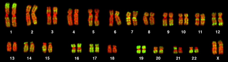
In a given species, chromosomes tin be identified past their number, size, centromere position, and banding pattern. In a man karyotype, autosomes or "body chromosomes" (all of the non–sex chromosomes) are generally organized in estimate order of size from largest (chromosome ane) to smallest (chromosome 22). The X and Y chromosomes are non autosomes. Still, chromosome 21 is actually shorter than chromosome 22. This was discovered afterward the naming of Down's syndrome equally trisomy 21, reflecting how this disease results from possessing one extra chromosome 21 (three total). Non wanting to change the name of this of import affliction, chromosome 21 retained its numbering, despite describing the shortest set of chromosomes. The chromosome "arms" projecting from either cease of the centromere may be designated as short or long, depending on their relative lengths. The short arm is abbreviated p (for "petite"), whereas the long arm is abbreviated q (because it follows "p" alphabetically). Each arm is farther subdivided and denoted by a number. Using this naming system, locations on chromosomes can be described consistently in the scientific literature.
Effort It
Although Mendel is referred to equally the "begetter of mod genetics," he performed his experiments with none of the tools that the geneticists of today routinely employ. Ane such powerful cytological technique is karyotyping, a method in which traits characterized by chromosomal abnormalities can be identified from a single cell. To discover an individual's karyotype, a person's cells (like white blood cells) are first collected from a blood sample or other tissue. In the laboratory, the isolated cells are stimulated to begin actively dividing. A chemical chosen colchicine is then applied to cells to arrest condensed chromosomes in metaphase. Cells are so fabricated to cracking using a hypotonic solution so the chromosomes spread apart. Finally, the sample is preserved in a fixative and applied to a slide.
The geneticist then stains chromosomes with one of several dyes to better visualize the singled-out and reproducible banding patterns of each chromosome pair. Following staining, the chromosomes are viewed using brilliant-field microscopy. A common stain choice is the Giemsa stain. Giemsa staining results in approximately 400–800 bands (of tightly coiled DNA and condensed proteins) arranged along all of the 23 chromosome pairs; an experienced geneticist can identify each band. In addition to the banding patterns, chromosomes are further identified on the basis of size and centromere location. To obtain the classic depiction of the karyotype in which homologous pairs of chromosomes are aligned in numerical order from longest to shortest, the geneticist obtains a digital prototype, identifies each chromosome, and manually arranges the chromosomes into this pattern (Figure 1).
At its nearly basic, the karyogram may reveal genetic abnormalities in which an private has too many or too few chromosomes per prison cell. Examples of this are Downward Syndrome, which is identified past a 3rd copy of chromosome 21, and Turner Syndrome, which is characterized by the presence of just one X chromosome in women instead of the normal two. Geneticists can also identify large deletions or insertions of Dna. For instance, Jacobsen Syndrome—which involves distinctive facial features as well as heart and bleeding defects—is identified by a deletion on chromosome 11. Finally, the karyotype tin can pinpoint translocations, which occur when a segment of genetic cloth breaks from ane chromosome and reattaches to another chromosome. Translocations are implicated in certain cancers, including chronic myelogenous leukemia.
During Mendel's lifetime, inheritance was an abstract concept that could only be inferred by performing crosses and observing the traits expressed by offspring. By observing a karyogram, today'south geneticists tin can actually visualize the chromosomal composition of an individual to confirm or predict genetic abnormalities in offspring, even earlier birth.
Common Disorders
Of all of the chromosomal disorders, abnormalities in chromosome number are the most obviously identifiable from a karyogram. Disorders of chromosome number include the duplication or loss of entire chromosomes, every bit well equally changes in the number of complete sets of chromosomes. They are caused by nondisjunction, which occurs when pairs of homologous chromosomes or sister chromatids fail to carve up during meiosis. Misaligned or incomplete synapsis, or a dysfunction of the spindle apparatus that facilitates chromosome migration, can cause nondisjunction. The risk of nondisjunction occurring increases with the historic period of the parents.
Nondisjunction tin occur during either meiosis I or Ii, with differing results (Figure ii). If homologous chromosomes fail to separate during meiosis I, the result is two gametes that lack that particular chromosome and ii gametes with two copies of the chromosome. If sister chromatids neglect to separate during meiosis II, the consequence is one gamete that lacks that chromosome, two normal gametes with one copy of the chromosome, and ane gamete with two copies of the chromosome.
Exercise Question

Which of the following statements about nondisjunction is true?
- Nondisjunction only results in gametes with n+one or northward–one chromosomes.
- Nondisjunction occurring during meiosis II results in 50 percent normal gametes.
- Nondisjunction during meiosis I results in l per centum normal gametes.
- Nondisjunction always results in 4 different kinds of gametes.
- Show Answer
-
Answer b is true.
Aneuploidy

An individual with the advisable number of chromosomes for their species is chosen euploid; in humans, euploidy corresponds to 22 pairs of autosomes and one pair of sexual practice chromosomes. An individual with an fault in chromosome number is described equally aneuploid, a term that includes monosomy (loss of one chromosome) or trisomy (gain of an extraneous chromosome). Monosomic human zygotes missing whatsoever one copy of an autosome invariably fail to develop to birth because they lack essential genes. This underscores the importance of "factor dosage" in humans. Well-nigh autosomal trisomies also fail to develop to nascence; however, duplications of some of the smaller chromosomes (13, xv, 18, 21, or 22) tin consequence in offspring that survive for several weeks to many years. Trisomic individuals suffer from a different type of genetic imbalance: an backlog in gene dose. Individuals with an actress chromosome may synthesize an abundance of the gene products encoded past that chromosome. This extra dose (150 percent) of specific genes can lead to a number of functional challenges and often precludes development. The most mutual trisomy amongst viable births is that of chromosome 21, which corresponds to Downwardly Syndrome. Individuals with this inherited disorder are characterized by curt stature and stunted digits, facial distinctions that include a broad skull and large tongue, and significant developmental delays. The incidence of Downward syndrome is correlated with maternal age; older women are more likely to become pregnant with fetuses carrying the trisomy 21 genotype (Figure three).
Polyploidy

An individual with more the right number of chromosome sets (ii for diploid species) is called polyploid. For case, fertilization of an abnormal diploid egg with a normal haploid sperm would yield a triploid zygote. Polyploid animals are extremely rare, with just a few examples amidst the flatworms, crustaceans, amphibians, fish, and lizards. Polyploid animals are sterile considering meiosis cannot proceed normally and instead produces generally aneuploid girl cells that cannot yield viable zygotes. Rarely, polyploid animals can reproduce asexually by haplodiploidy, in which an unfertilized egg divides mitotically to produce offspring. In contrast, polyploidy is very common in the plant kingdom, and polyploid plants tend to be larger and more robust than euploids of their species (Effigy 4).
Sex Chromosome Nondisjunction in Humans
Humans display dramatic deleterious furnishings with autosomal trisomies and monosomies. Therefore, information technology may seem counterintuitive that human females and males tin office normally, despite conveying different numbers of the X chromosome. Rather than a gain or loss of autosomes, variations in the number of sex chromosomes are associated with relatively mild effects. In part, this occurs considering of a molecular process called X inactivation. Early on in development, when female mammalian embryos consist of just a few thousand cells (relative to trillions in the newborn), i Ten chromosome in each jail cell inactivates by tightly condensing into a quiescent (dormant) structure called a Barr trunk. The chance that an X chromosome (maternally or paternally derived) is inactivated in each prison cell is random, but one time the inactivation occurs, all cells derived from that ane will have the same inactive Ten chromosome or Barr body. Past this process, females compensate for their double genetic dose of X chromosome.

In so-called "tortoiseshell" cats, embryonic Ten inactivation is observed every bit color variegation (Figure 5). Females that are heterozygous for an Ten-linked glaze color cistron will express one of two different coat colors over unlike regions of their body, respective to whichever 10 chromosome is inactivated in the embryonic jail cell progenitor of that region.
An individual carrying an abnormal number of X chromosomes will inactivate all but one 10 chromosome in each of her cells. Still, even inactivated Ten chromosomes continue to express a few genes, and X chromosomes must reactivate for the proper maturation of female ovaries. As a result, X-chromosomal abnormalities are typically associated with mild mental and physical defects, as well as sterility. If the X chromosome is absent altogether, the private will not develop in utero.
Several errors in sexual activity chromosome number have been characterized. Individuals with three X chromosomes, called triplo-10, are phenotypically female person only express developmental delays and reduced fertility. The XXY genotype, respective to ane type of Klinefelter syndrome, corresponds to phenotypically male person individuals with pocket-size testes, enlarged breasts, and reduced body hair. More complex types of Klinefelter syndrome exist in which the individual has equally many as five Ten chromosomes. In all types, every X chromosome except ane undergoes inactivation to compensate for the excess genetic dosage. This can be seen equally several Barr bodies in each cell nucleus. Turner syndrome, characterized as an X0 genotype (i.east., only a single sexual activity chromosome), corresponds to a phenotypically female individual with brusque stature, webbed skin in the cervix region, hearing and cardiac impairments, and sterility.
Duplications and Deletions
In addition to the loss or proceeds of an unabridged chromosome, a chromosomal segment may be duplicated or lost. Duplications and deletions often produce offspring that survive but showroom physical and mental abnormalities. Duplicated chromosomal segments may fuse to existing chromosomes or may be gratuitous in the nucleus. Cri-du-chat (from the French for "cry of the cat") is a syndrome associated with nervous system abnormalities and identifiable physical features that result from a deletion of most of 5p (the small arm of chromosome 5) (Effigy half dozen). Infants with this genotype emit a feature high-pitched weep on which the disorder's name is based.

Check Your Understanding
Respond the question(southward) below to meet how well you empathise the topics covered in the previous section. This short quiz does not count toward your grade in the form, and yous tin can retake information technology an unlimited number of times.
Use this quiz to check your understanding and decide whether to (1) study the previous section further or (2) move on to the adjacent section.
https://assessments.lumenlearning.co...sessments/6890
Source: https://bio.libretexts.org/Courses/Lumen_Learning/Book:_Biology_for_Non-Majors_I_%28Lumen%29/07:_Cell_Division/7.08:_Errors_in_Chromosome_Number
Posted by: maysracion.blogspot.com
0 Response to "What Is A Question That Could Be Answered By Observing Chromosomes Of Different Species Of Animals"
Post a Comment